Microscopy VS PCR
Airborne spore trapping is an essential method for monitoring the risks of fungal diseases in agriculture. Two main approaches exist to identify and count spores: microscopy and quantitative PCR (qPCR). While PCR is often considered the more modern technique, microscopy offers several concrete advantages, especially in the context of field prevention.
1. Direct and Rapid Observation
Microscopy allows for immediate analysis of collected spores, without complex DNA extraction steps or the need for a specialized laboratory. In crop disease prevention, the speed of reporting is a critical factor. Once contamination is detected, it is essential to act quickly with preventive or curative treatments before the disease spreads and severely affects the crop.
With PCR, analyses are usually centralized and must be shipped to specialized labs, which often leads to delays of several days before results are available. This makes it difficult to intervene on time. In contrast, microscopy is a portable method that can be performed near the sampling site, enabling much faster reaction times and maximizing the chances of protecting crops before it is too late.
2. Sensitivity
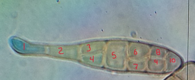
The sensitivity of a method refers to its ability to detect true positives when they are present. For spores with distinct morphological traits, microscopy and PCR have theoretically comparable sensitivity. Microscopy can detect a disease as soon as a single spore (or sporangium) is present in the sample. Similarly, PCR should detect a single spore if the reaction runs properly.
When comparing quantitative PCR results with microscopy counts for the same sample, PCR often appears more sensitive because genome counts per liter of air are typically higher or equal to spore counts. However, this difference arises because several fungal pathogens, such as Alternaria and Phytophthora, contain multiple genomes per spore. For example, 10 genomes detected by PCR or one spore observed under the microscope are equivalent and correspond to capturing a single spore.
Ultimately, the sensitivity of both methods depends more on the air sampling strategy and timing than on the analysis method itself.
3. Specificity
Specificity is the ability to correctly identify true negatives. With microscopy, distinguishing between certain pathogenic species and closely related species can sometimes be challenging. However, our comparative studies with PCR show that, in agricultural field sampling, our microscopy method correctly identifies the targeted pathogens over 95% of the time.
4. Method Validation and Execution
Both PCR and microscopy require solid validation processes to ensure optimal sensitivity. For microscopy, this involves studying spore varieties that are morphologically similar to the target spores and identifying discriminating traits for reliable identification. Microscope slide preparation is non-destructive and does not hinder spore detection.
For PCR, validation is equally crucial, but since the method involves multiple sequential steps, an error at any stage can drastically affect the results. Another advantage of microscopy is that each slide can be reanalyzed immediately by anyone, using the original sample. In contrast, qPCR requires a complete new sample preparation for reanalysis.
5. Broad View and Diversity
Unlike PCR, which targets specific species using primers, microscopy allows for the observation of all spores present in the sample, including non-targeted or emerging pathogens. This broader perspective provides valuable insights into the phytosanitary risks of a field or region.
PCR remains a valuable method for specific confirmations or targeted research where precise genetic identification is required. However, for routine surveillance, microscopy stands out as the preferred option due to its unmatched speed and high reliability. It enables early and comprehensive detection of airborne pathogens, making it an ideal tool for the daily monitoring of fungal diseases in agriculture.